Don't just leave it to a gut feeling - find out how to interpret abdominal radiographs properly
Abdominal Radiology
Mini Series
Get all the practical help you need to evaluate abdominal radiographs in this easy to attend, fully guaranteed, Online Mini Series™ from CPD Solutions
- Learn how to recognise abnormalities
- You have a whole year’s access to recorded sessions and study notes for reviewing key points
- Superb value for money – you learn without travelling or paying for accommodation, childcare or petcare
- Watch the recordings on your iPad, mobile, PC or tablet
- Self-assessment quiz to ‘release’ your 8 hours CPD certification
This course will help me in practice a great deal – improved confidence in interpretation, more likely to pick up on abnormalities that would previously overlooked
This course was very informative and helped me gain confidence in reading radiographs as a new graduate!
What will I learn on this course?
Session 1
Getting the Best out of Your Abdominal Radiographs
- Obtaining diagnostic abdominal radiographs – what are the potential challenges?
- Principles of interpretation – remember Roentgen signs?
- Abdominal radiology vs ultrasound – do I need both?
- A logical approach to abdominal masses
- Normal radiographic anatomy of the peritoneal and retroperitoneal spaces, identifying free fluid and free gas, typical radiographic features of peritonitis and abdominal lymphadenopathy
- Recognising changes in hepatic size, shape and opacity, typical radiographic features of liver masses, metabolic hepatopathy, congenital porto-systemic shunts and chronic liver disease
- Recognising changes in splenic size, shape and opacity, typical radiographic features of splenic masses and splenic torsion
- What could ultrasound add to the evaluation of the abdominal space, liver and spleen?
- Test yourself: Examples of commonly encountered hepatic, splenic and abdominal space disease
Session 2
Evaluating the Uro-genital Tract
- Evaluating the ovaries, uterus and prostate
- Radiographic features of pregnancy, pyometra, uterine and ovarian neoplasia
- Radiographic features of prostatic disease, paraprostatic cysts and cryptorchidism
- What could ultrasound add to the evaluation of the urogenital tract?
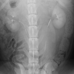
- Evaluating the kidneys, ureters, bladder and urethra
- Recognising changes in renal size, shape and opacity
- Urinary tract contrast studies
- Radiographic features of renal & ureteral calculi, nephritis / pyelonephritis, hydronephrosis, ectopic ureters, renal neoplasia, renal cysts and renal and/or ureteral rupture
- Radiographic features of cystitis, bladder and urethral calculi, bladder and urethral neoplasia, bladder and urethral rupture and bladder diverticula
Session 3
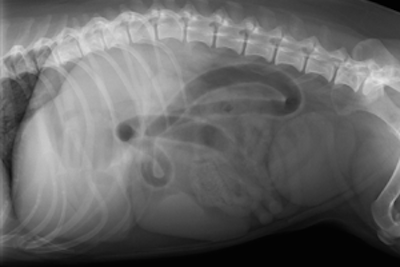
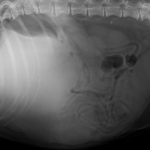
Evaluating the Gastro-intestinal Tract
- Evaluating the stomach and radiographic anatomy and variations
- Identifying gastric foreign bodies
- Typical radiographic features of gastric dilation and volvulus
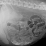
- Evaluating the small intestine
- Logical evaluation of changes in the size, shape, opacity and position if the small intestinal loops
- Recognising small intestinal obstruction
- Can I recognise pancreatitis from an abdominal radiograph?
- Evaluating the large intestine
- Typical radiographic features of ileo-caeco-colic intussusceptions, constipation, mega-colon and perineal hernia
- When and how should I perform gastro-intestinal contrast studies?
- What could ultrasound add to the evaluation of the gastro-intestinal tract and pancreas?
- Test yourself: Examples of commonly encountered gastro-intestinal abnormalities
Meet the Presenter....
Esther Barrett
MA VetMB DVDI DipECVDI MRCVS
RCVS & European Specialist in Veterinary Diagnostic Imaging

Esther spent 6 years working in mixed first opinion practice in the UK, before taking up a residency in Veterinary Diagnostic Imaging. She obtained the RCVS and European College Diplomas in Veterinary Diagnostic Imaging in 2006, and after spending 2 further years working in University referral practice, she set up her own freelance veterinary imaging service.
As well as loving the practical challenges of her day-to-day work, Esther also enjoys teaching: she regularly provides small group CPD for veterinary surgeons throughout the UK and in 2010 she was appointed as a Special Lecturer in Veterinary Diagnostic Imaging for the University of Nottingham.
Watch the recordings at a time convenient to you!
Prior to this course I hadn’t had much experience with thoracic radiographs and they all looked the same to me, so this has been very helpful in helping me know what I am looking at.
12 months access to recordings and course materials is included
Please note that these are webinar recordings and not live events.
Full details on how to access the Mini Series will be emailed to you.

Take advantage of the easy Online Mini Series™ and develop a logical approach to interpreting abdominal radiographs
Just £447 + VAT*
Overseas customers outside the UK will not be charged VAT.

Price includes...
- All 3 sessions
- Notes and quiz (8 hours CPD)
- PLUS unlimited access to recordings and all course materials for 12 months!
- No traffic jams or accommodation hassles
- No child or pet care to arrange
- No rota clashes to worry about and no locum cover needed
Just great CPD and a valuable ongoing resource

100% Money Back Guarantee
Buy with absolute confidence as your purchase is protected by our 100%, no quibble money-back guarantee
Film reading and good comprehensive course notes most useful. Course will help with interpreting abdominal x-rays in practice, in particular suspect FB.

Need Some Help?
Have A Question?
Please call us on:
0151 328 0444Alternatively email
webinarclub@cpd-solutions.comWe will be delighted to help. Thank you!